Hsv Esophagitis Punched Out Ulcers : Gastroesophageal Reflux Disease Amboss - Candida, hsv1 (punched out lesions), cmv (linear lesions) eosinophilic esophagitis:. Esophagitis, also spelled oesophagitis, is a disease characterized by inflammation of the esophagus. Other pathogens include cytomegalovirus (cmv), herpes simples virus (hsv) and hiv (idiopathic) esophagitis may be the cause if no other pathogens are found. Cmv esophagitis linear ulcers owl's eye cells. Symptoms include pain or a burning sensation behind or below the sternum, the flat bone that runs down the center of the chest. Esophagitis can cause painful, difficult swallowing and chest pain.
Discrete ulcers or ulcers with vesicles, bullae, or pseudomembranes. Treatment for esophagitis depends on the underlying cause and the severity of tissue damage. Swallowed food and liquids normally pass through it. Cmv esophagitis linear ulcers owl's eye cells. This can help you and your doctor figure out which foods might cause your esophagitis.
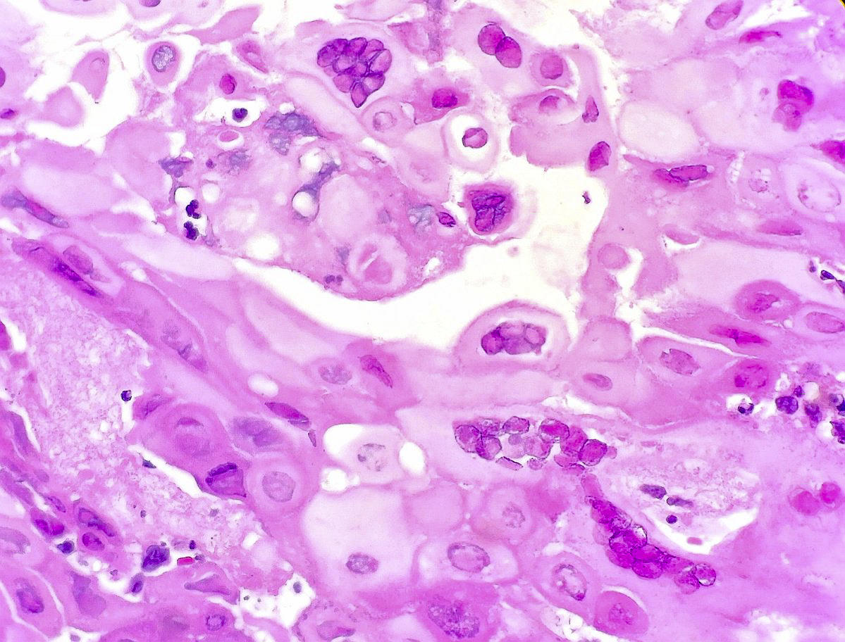
Pathology Outlines Herpes Simplex Esophagitis from www.pathologyoutlines.com Other pathogens include cytomegalovirus (cmv), herpes simples virus (hsv) and hiv (idiopathic) esophagitis may be the cause if no other pathogens are found. Grossly, hsv esophagitis shows erosive changes to the mucosa and frequently multiple superficial ulcers. Must rule out hsv infection as cause of esophageal ulcers, particularly from immunocompromised patients. Causes of esophagitis include stomach acids backing up into the esophagus, infection, oral medications and allergies. Hsv esophagitis is the second most common cause of infectious esophagitis and it occurs in both immunocompromised and immunocompetent patients. Histology is rather nonspecific, with erosions. Differential diagnosis between herpes simplex virus (hsv) esophagitis and cytomegalovirus (cmv) esop. You'll see rings in the esophagus and biopsy will show eosinophils.
Esophagitis is when the lining of your esophagus becomes irritated and inflamed.
Esophagitis can cause painful, difficult swallowing and chest pain. Cmv esophagitis linear ulcers owl's eye cells. Swallowed food and liquids normally pass through it. It connects the pharynx to the stomach; Candida wont present with a visible ulcer, it'll have candidial growth on top(duh! Herpesvirus (hsv) esophagitis is seen predominantly in patients who are immunocompromised. Solid organ and bone marrow transplant. Occasionally, herpes esophagitis manifests as multiple plaquelike lesions in the esophagus, but this infection is more commonly associated with small, superficial ulcers (see herpes esophagitis, below). Hsv shows punched out ulcers in esophagus. Differential diagnosis between herpes simplex virus (hsv) esophagitis and cytomegalovirus (cmv) esophagitis is challenging the endoscopic features of esophagitis were categorized and scored as follows: „ odynophagia is the predominant presenting symptom. Histology is rather nonspecific, with erosions. Herpes esophagitis is an inflammation of the esophagus due to herpes simplex virus.
„ odynophagia is the predominant presenting symptom. If you have a health problem or are having treatment that weakens your immune system. People with a normal immune system are not likely to get infectious esophagitis. Think of this in atopic patients, like those with asthma, eczema or food allergies. Biopsies should be taken from both the center and periphery of the ulcer.
Histologic Section Of An Esophageal Biopsy Obtained From A 36 Year Old Download Scientific Diagram from www.researchgate.net Discrete ulcers or ulcers with vesicles, bullae, or pseudomembranes. Hsv esophagitis is the second most common cause of infectious esophagitis and it occurs in both immunocompromised and immunocompetent patients. Esophagitis can cause painful, difficult swallowing and chest pain. Were more frequent in patients with hsv esophagitis, whereas. People with a normal immune system are not likely to get infectious esophagitis. Infectious esophagitis in the immunocompetant. Pylori or chronic nsaid use usually in superior duodenum or pylorus of the stomach perforation, obstruction, and anemia can result. „ odynophagia is the predominant presenting symptom.
Ulcers can coalese and form black esophagus histology details:
Treatment for esophagitis depends on the underlying cause and the severity of tissue damage. Symptoms include pain or a burning sensation behind or below the sternum, the flat bone that runs down the center of the chest. Hsv esophagitis punched out ulcer tzanck test for syncitia. Think of this in atopic patients, like those with asthma, eczema or food allergies. Candida and herpes simplex virus (hsv) esophagitis has been reported in 2% to 4% in immunocompromised patients. Esophageal ulcers are a type of peptic ulcer that develops between the throat and the stomach. Discrete ulcers or ulcers with vesicles, bullae, or pseudomembranes. Swallowed food and liquids normally pass through it. Who is at risk for infectious esophagitis? Esophagitis is when the lining of your esophagus becomes irritated and inflamed. Although hsv esophagitis is much more common in immunosuppressed individuals, it can occur in healthy persons. This can help you and your doctor figure out which foods might cause your esophagitis. Histology is rather nonspecific, with erosions.
This can help you and your doctor figure out which foods might cause your esophagitis. Hsv esophagitis punched out ulcer tzanck test for syncitia. Candida and herpes simplex virus (hsv) esophagitis has been reported in 2% to 4% in immunocompromised patients. Viral culture if no treatment response. Solid organ and bone marrow transplant.
Pin On Medicina from i.pinimg.com 38 year old woman with esophagus ulcer; „ odynophagia is the predominant presenting symptom. Herpes esophagitis is a viral infection of the esophagus caused by herpes simplex virus (hsv). Hsv esophagitis punched out ulcer tzanck test for syncitia. Candida, hsv1 (punched out lesions), cmv (linear lesions) eosinophilic esophagitis: Ulcers can coalese and form black esophagus histology details: Were more frequent in patients with hsv esophagitis, whereas. You'll see rings in the esophagus and biopsy will show eosinophils.
Occasionally, herpes esophagitis manifests as multiple plaquelike lesions in the esophagus, but this infection is more commonly associated with small, superficial ulcers (see herpes esophagitis, below).
Herpes esophagitis is a viral infection of the esophagus caused by herpes simplex virus (hsv). Histology is rather nonspecific, with erosions. Differential diagnosis between herpes simplex virus (hsv) esophagitis and cytomegalovirus (cmv) esophagitis is challenging the endoscopic features of esophagitis were categorized and scored as follows: Must rule out hsv infection as cause of esophageal ulcers, particularly from immunocompromised patients. Candida and herpes simplex virus (hsv) esophagitis has been reported in 2% to 4% in immunocompromised patients. Solid organ and bone marrow transplant. Using medications, bodily infections, and exposure to stomach acid are. Hsv esophagitis is the second most common cause of infectious esophagitis and it occurs in both immunocompromised and immunocompetent patients. Biopsies should be taken from both the center and periphery of the ulcer. It connects the pharynx to the stomach; Hsv shows punched out ulcers in esophagus. Although hsv esophagitis is much more common in immunosuppressed individuals, it can occur in healthy persons. Esophageal ulcers are a type of peptic ulcer that develops between the throat and the stomach.
Large ulcers (red, black and white arrows) are seen in these two views of the distal esophagus from hsv esophagitis. Were more frequent in patients with hsv esophagitis, whereas.


Komentar
Posting Komentar